Case Study | 2. Excruciating Back Pain
A patient was offered painkillers and surgery to fix her debilitating back pain, which her doctors told her was caused by a herniated disc. After reviewing her case, we solved it for $7.

A Bit of Context
One evening, my father asked me if I would review the case of a close family friend who is suffering from worsening back pain. This person lives in the developing country from which I emigrated, where access to robust radiology services are limited.
Of course, I agreed to take a look if only to provide confidence in their diagnosis.
How optimistic.
This is a particularly fascinating case because it illustrates:
Inadequacy of standardized medical training
Inability of licensed professionals to demonstrate insight
Limitations of high-quality imaging to provide answers
How pills & surgery can be avoided with critical thought
Case History
The client was a middle-age woman with a new onset of low back pain, which had gradually progressed and now prevented her from going about her daily life. She could barely get out of bed.
Her workup included the usual diagnostic testing, including x-rays and an MRI of her lumbar spine.
At the end of her workup, the doctors concluded that she had some mild degenerative disease of her lumbar spine with small disc herniations.
She was offered the standard combination of painkillers and possible surgical intervention.
The Second Opinion
Shortly after the conclusion of the workup, I was sent all her imaging and asked for my opinion.
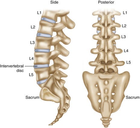
I began by looking at the radiologist’s report, then compared to the images. The report mentioned relatively small herniations at 3 contiguous intervertebral disc levels: between L3-L4, L4-L5 and L5-S1. Scrolling through the images, I could see why the radiologist read the study as they did.
The report was a classic description of lumbar spine anatomy & degeneration. This is precisely how most radiologists are trained to describe the findings of a Lumbar Spine MRI.
Looking at simply the imaging findings and the patient’s history, the standard fiat physician would not be held at fault for interpreting the MRI as they did.
However, as we look closer and think more deeply of spinal biomechanics, we begin to appreciate how much was missed!


